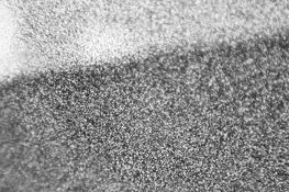
viewing emulsion under a microscope

Recent Classifieds

Forum statistics

Threads
197,580
Messages
2,761,331
Members
99,406
Latest member
filmtested
Recent bookmarks
0

rmazzullo

Subscriber
Joined
Mar 24, 2007
Messages
321
Location
Northeast US
Format
Multi Format
PE,

Is there a way of using an optical microscope to examine grain size, distribution and shape? Do you have any thoughts on magnification and / or microscope type? How would the emulsion sample be prepared?

Thanks,

Bob M.
 

ben-s

Member
Joined
Jul 12, 2006
Messages
444
Location
Nottingham,
Format
Multi Format
I've had developed emulsions under a microscope:
2939977040_7fa099d9a7.jpg

http://www.flickr.com/photos/ben-s/2939977040/
That was with a 10x objective, no eyepiece and a home made camera adapter.

I've also tried looking side-on at emulsions under the microscope. I found it helped to swell the emulsion first, otherwise it's all base and a tiny layer of emulsion on top.
The most difficult bit was cutting the edge accurately enough, as I don't have a microtome.
 

MikeSeb

Subscriber
Joined
Jun 12, 2005
Messages
1,104
Location
Denver, CO
Format
Medium Format
Bob, without wanting to seem snarky--what is the purpose for doing this? I mean, I'm no stranger to purposeless activities myself ( :smile: ) but what does this achieve?

Best holiday wishes.
 
OP
OP
rmazzullo

rmazzullo

Subscriber
Joined
Mar 24, 2007
Messages
321
Location
Northeast US
Format
Multi Format
Hello Mike, Ben....

Ben,

A long time ago, I remember seeing an article where the writer made a microtome from a 3/8 - 1/2 inch (10 - 12 mm thereabouts) diameter nut and bolt. He rotated the nut to almost the end of the bolt (before it would normally fall off) and embedded the object in epoxy (or wax, I think) in the open space. Once the material hardened, he rotated the nut onto the bolt in very small increments to expose the embedded material, and sliced off what he needed. I will have to draw a diagram and scan it in later on.

Mike,

This is a good question, and I don't mind at all.....

The reason I am asking about the microscope is that I believe it would be helpful if you could actually see what kind of grains (size, shape, etc) result from the different emulsion types and formulas that are possible - when you make them in your location. Even if the formulas are followed exactly, there are different factors in play that can change things. Do you have hard or soft water? Other elements in the water? Copper pipes or PVC or cast iron? Is your thermometer / thermostat / heating element calibrated? What are you using for a timer? Addition rates and methods over time? Things like that. I would imagine that it would be very revealing if you could actually see what kind of grains result from a particular emulsion making setup and procedure.

Thanks, and have a great holiday!

Bob M.
 
Last edited by a moderator:

MikeSeb

Subscriber
Joined
Jun 12, 2005
Messages
1,104
Location
Denver, CO
Format
Medium Format
Well, duh, it makes perfect sense for an emulsion MAKER. I'm just an emulsion-exposer, so what would I know?

:wink:
 
OP
OP
rmazzullo

rmazzullo

Subscriber
Joined
Mar 24, 2007
Messages
321
Location
Northeast US
Format
Multi Format
Mike,

I could see where it might be interesting to see what you come up with after the emulsion is exposed and processed. I guess it depends on how far you want to take it.

Bob
 

c.d.ewen

Subscriber
Joined
Jan 27, 2005
Messages
685
Location
Northeast USA
Format
ULarge Format
Bob, without wanting to seem snarky--what is the purpose for doing this? I mean, I'm no stranger to purposeless activities myself ( :smile: ) but what does this achieve?

Best holiday wishes.

Mike:

Sorry to be in a snarky mode, but it's 1PM and the adults haven't opened their presents yet.

The purpose is to have an excuse to buy a [better] microscope :wink:

Best holiday wishes to all, and a Merry Christmas to those who need one.

Charley
 

Photo Engineer

Subscriber
Joined
Apr 19, 2005
Messages
29,018
Location
Rochester, NY
Format
Multi Format
Guys;

A microscope at about 2500X or higher with oil immersion if possible, will help one view layer composition and developed silver grains as well as dye droplets in color film. This will show gross structure for comparisons. We use this primarily for film design analysis. It is rather coarse and low resolution for anything other than this type of work.

For details and emulsion work, you have to use an electron microscope to see grains and you need something on the order of 0.1 - 10 micron scale to do this. The grain must be made into a carbon replica or the like and the gelatin must be removed to prevent obstruction of the image. These have to be done in specialized labs and are pretty pricey. The silver halide grains are outside of the range of a microscope except as featureless dark dots in most microscopic pictures.

Mastering the microtome method for film analysis is a painstaking job for a craftsman. The microtomed section must be mounted flat in order to see the object correctly. I worked on this for hours at a time, but was never really any good at it myself. We had an open lab for this type of work, but I ended up having to submit my work for a pro to do it.

As a side note, the guy who I usually worked with was a nephew of Anton Dvorak the famous composer. He was a resistance fighter during WWII and had some amazing stories to tell. He was a true artist with a microtome and with the microscope.

PE
 

Jim Noel

Member
Joined
Mar 6, 2005
Messages
2,261
Format
Large Format
PE beat me to the punch.
What you are seeing with an optical microscope is not the grains, but clusters of grain.
 
OP
OP
rmazzullo

rmazzullo

Subscriber
Joined
Mar 24, 2007
Messages
321
Location
Northeast US
Format
Multi Format
Damn...0 for 2. I'm on a streak!! :wink:

Thanks PE and everyone for their input. I guess I'll have to wait until an electron microscope comes up for sale on e-bay. (I'm just kidding).

Happy holidays to all,

Bob M.
 
Joined
Apr 18, 2005
Messages
818
Location
San Bernardi
Format
8x10 Format
HAPPY HOLIDAYS TO ALL EMULSION MAKERS !
There are many electron microscopes on ebay! Price ranges from under a thousand dollar to the mid 10s of thousands. I am gonna stick with my optical scopes. I have known people who had full time jobs just preparing samples for and running those things.
Bill
 

Photo Engineer

Subscriber
Joined
Apr 19, 2005
Messages
29,018
Location
Rochester, NY
Format
Multi Format
PE beat me to the punch.
What you are seeing with an optical microscope is not the grains, but clusters of grain.


With an optical microscope, you see a hint of the silver halide grain as translucent dots before development. After development you see black threads, clumps and dots of silver metal.

I forgot to mention that here, sample prep involves gluing the emulsion side of the film to a substrate very firmly before microtoming in order to prevent deformation or destruction of the emulsion layer. And, photmicrographs of raw emulsion are rather rare.

With an electron microscope, you see the crystal as it actually is.

Electron micrographs run from about $150 to 500 each depending on lab service offered and quantity done at one time.

Both types of imaging have been done on emulsions and pubilshed in textbooks, but electron micrographs are the preferred method for raw emulsion and photo micrographs are preferred for film cross sections.

PE
 
OP
OP
rmazzullo

rmazzullo

Subscriber
Joined
Mar 24, 2007
Messages
321
Location
Northeast US
Format
Multi Format

Photo Engineer

Subscriber
Joined
Apr 19, 2005
Messages
29,018
Location
Rochester, NY
Format
Multi Format
Bob;

This is not the same thing. See the photo quality images that I posted elsewhere at the 1 micron resolution.

PE
 
OP
OP
rmazzullo

rmazzullo

Subscriber
Joined
Mar 24, 2007
Messages
321
Location
Northeast US
Format
Multi Format
PE,

I should have mentioned that my post was meant more for it's novelty value than for any real practical use.

Oops.

Bob M.
 

David Foy

Member
Joined
Jul 20, 2004
Messages
6
Location
Calgary
Format
Multi Format
electron microscope

Scientific American published instructions for making an electron microscope at home. Martin Gardner's column, I believe. Check the indexes from 30 to 40 years ago. If I remember right, it involved little more than the kind of parts, equipment and glass work they use at neon sign places.

...just can case anyone is getting bored ...
 

Alan Johnson

Subscriber
Joined
Nov 16, 2004
Messages
3,224
My 1950's microscope is OK for making photomicrographs of edge effects and of negatives of resolution charts but grain(clumps) is a bit beyond it. Attachment,IIRC FP4 in Pyrocat HD with a 40x objective was hard to focus and dim.A modern microscope might do better.
 

Attachments

  • grain.jpg
    grain.jpg
    184.6 KB · Views: 244

Photo Engineer

Subscriber
Joined
Apr 19, 2005
Messages
29,018
Location
Rochester, NY
Format
Multi Format
Well, here are some things to consider when wanting to look at the internal structure of a film:

1. Looking at the raw emulison - use an Electron Microscope or a Scanning Electron Microscope. Sample prep is critical to see the grains, and resolution must be on the order of 1.0 micron or smaller in the case of paper emulsions. Don't forget the words "SAMPLE PREP". These were done in a special lab. I did quite a few, but never ran the instrument.

2. A microdensitometer is used to look at edge effects. I have posted examples here on APUG. I did a lot of these, but never ran the instrument.

3. Grain is measured as RMSG (Root Mean Square). To do this a microdensitometer is used with special software / hardware to derive the grain profile. A sample is scanned across a given step to get the RMS of the deviation in density. I have no examples, but used to do this often. I never ran this instrument.

4. To look at film structure, you need a microtome and a method of laminating the film to another substrate. You cut sandwiches of this and look edge on to see the structure. Silver grains and silver can be seen as tiny dots with no resolution. Usually, the magnifiacation is about 2500x and an oil immersion lens is used. I did a lot of these myself but was never what you would consider an expert.

PE
 

Photo Engineer

Subscriber
Joined
Apr 19, 2005
Messages
29,018
Location
Rochester, NY
Format
Multi Format
Basically a microdensitometer is a regular densitometer recalibrated to have a high intensity beam with a small aperture. It must be smaller than the smallest line you wish to scan. We typically used 1000, 100, 10 and 1 micron lines.

Usually, 10 microns is small enough, and therefore a 1 micron or 0.5 micron aperture would be OK.

PE
 

Photo Engineer

Subscriber
Joined
Apr 19, 2005
Messages
29,018
Location
Rochester, NY
Format
Multi Format
Grain clumps are smaller, and appear as noise in photomicrographs of edges. This is how RMSG is calculated, by analyzing the noise.

PE
 

archphoto

Member
Joined
Dec 14, 2008
Messages
960
Location
Holland and
Format
4x5 Format
By all means get yourself a better microscope !
The type depends on what you want to see: butterfly wings can be seen with low magnification, filmgrain at verry high.
I know this is a bit on the side-line of this forum, but still, you can allways send me a personal message.

Good luck !
Peter
 

patrickjames

Member
Joined
Mar 25, 2005
Messages
742
Format
Multi Format
This is a little off topic, but about 15 years ago I know a rather brilliant fellow who was making an electron microscope the size of a sugar cube as his doctoral thesis. At the time it blew my mind that it was so small. I can only imagine the size they can make them now!
 
Photrio.com contains affiliate links to products. We may receive a commission for purchases made through these links.
To read our full affiliate disclosure statement please click Here.

PHOTRIO PARTNERS EQUALLY FUNDING OUR COMMUNITY:


Ilford ADOX Freestyle Photographic Stearman Press Weldon Color Lab Blue Moon Camera & Machine
Top Bottom